Mostrar el registro sencillo del ítem
Evaluation of Three Models of Follow-up of Patients with Cervical Cytological Abnormalities in a Peruvian’s Largest Teaching Women’s Hospital
| dc.contributor.author | Moya-Salazar, Jeel | es_ES |
| dc.contributor.author | Huarcaya, Jennifer | es_ES |
| dc.contributor.author | Rojas-Zumaran, Víctor | es_ES |
| dc.contributor.author | Vásquez, Diana L. | es_ES |
| dc.contributor.author | Chicoma-Flores, Karina | es_ES |
| dc.contributor.author | Contreras-Pulache, Hans | es_ES |
| dc.date.accessioned | 2022-10-10T21:48:01Z | |
| dc.date.available | 2022-10-10T21:48:01Z | |
| dc.date.issued | 2022-01-13 | |
| dc.identifier.uri | https://hdl.handle.net/20.500.13053/6852 | |
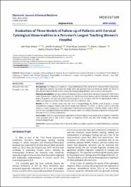
| dc.description.abstract | Introduction: The follow-up of squamous intraepithelial lesions (SIL) allows us to understand their progression and regression, however squamous cell atypia (ASC) can generate confusing follow-up results. We aimed to describe the evolution of ASC and SIL during cyto-histopathological follow-up in a tertiary-care hospital. Materials and methods: we conducted a retrospective study during 2016 in 156 Papanicolaou test (PAP) results under three models: 1) with ≥1 PAP and biopsies, 2) 1 PAP followed by ≥1 biopsy, and 3) ≥1 PAP and a confirmatory biopsy. Progression was defined as ASCUS to low-grade SIL (LSIL) or higher, and LSIL to high-grade SIL (HSIL) or higher; and regression as HSIL to LSIL or lower; and LSIL to ASCUS or lower. Results: In PAP, 57 (36.5%) cases were ASC and in histopathology 56 (39.9%) cases of grade 1 cervical intraepithelial neoplasia. Twenty-nine (18.6%) results were followed: 8 (27.6%), 17 (58.6%), and 4 (13.8%) with models 1, 2, and 3, respectively. The progression of the lesions was reported in ~50% for models 2 and 3. ASCUS was the main cytological finding that indicated biopsies, and for all models, the mean progression and regression time was 4 and 3.1 months, respectively. Conclusions: The follow-up of cytological alterations in three models showed progression of lesions in half of the cases analyzed with a time of four months of evolution; ASCUS was the main finding that indicated histopathological study. | es_ES |
| dc.format | application/pdf | es_ES |
| dc.language.iso | eng | es_ES |
| dc.publisher | Modestum LTD | es_ES |
| dc.rights | info:eu-repo/semantics/openAccess | es_ES |
| dc.rights.uri | https://creativecommons.org/licenses/by/4.0/ | es_ES |
| dc.subject | atypical squamous cells of the cervix, follow-up studies, squamous intraepithelial lesions, cytology, Peru | es_ES |
| dc.title | Evaluation of Three Models of Follow-up of Patients with Cervical Cytological Abnormalities in a Peruvian’s Largest Teaching Women’s Hospital | es_ES |
| dc.type | info:eu-repo/semantics/article | es_ES |
| dc.identifier.doi | https://doi.org/10.29333/ejgm/11546 | es_ES |
| dc.type.version | info:eu-repo/semantics/publishedVersion | es_ES |
| dc.publisher.country | GB | es_ES |
| dc.subject.ocde | http://purl.org/pe-repo/ocde/ford#3.03.00 | es_ES |
Ficheros en el ítem
Este ítem aparece en la(s) siguiente(s) colección(es)
-
SCOPUS [380]


